
Myelin is far more than insulation. The fatty sheaths that oligodendrocytes wrap around axons don't just speed signals along; they also help tune the timing of neural circuits, support the metabolic needs of neurons, and remodel as we learn. Yet for a structure first described nearly a century ago, we still understand remarkably little about how myelin is actually built.
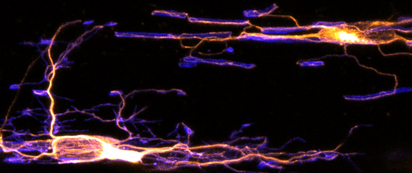
Textbook models hold that an oligodendrocyte contacts an axon and wraps it steadily from the outside in. By following individual sheaths as they formed, we found this isn't quite right: wrapping happens asynchronously, leaving partially wrapped zones that let a growing process reach past axon branches and link neighboring sheaths together by thin cytoplasmic bridges. This flexibility lets a single cell myelinate more of a circuit than we thought possible, but it leaves the resulting chains of sheaths vulnerable to degeneration with age.
That finding — a structure no existing model predicted — is what motivates the questions below. Read the full story on our Publications page
Every oligodendrocyte begins as a progenitor faced with a dramatic decision: whether to self-renew or differentiate. When it commits to differentiation, it rewrites nearly its entire genetic program in a day. In the mammalian brain, this attempt fails as often as it succeeds, ending in the cell's death. We want to know what orchestrates this rapid transformation. Surprisingly, part of the answer appears to lie in genes best known for driving cell division, repurposed here not to divide a cell, but to build myelin.
Using a targeted CRISPR-Cas9 screen in zebrafish, we identified several cyclin and cyclin-dependent kinase (CDK) genes required for oligodendrocyte generation — an unexpected role for machinery classically tied to the cell cycle. We're focused on cyclin Y-like 1 (CCNYL1): mutants show reduced oligodendrogenesis with no change in progenitor numbers, pointing to a specific defect in differentiation rather than proliferation. CCNYL1 is thought to act through CDK16 to regulate Wnt signaling, offering a fresh handle on a notoriously tangled pathway in this lineage. We're now defining its molecular partners in oligodendrocytes and using conditional mouse models to ask how these regulators matter across the lifespan throughout development, demyelination, and aging.
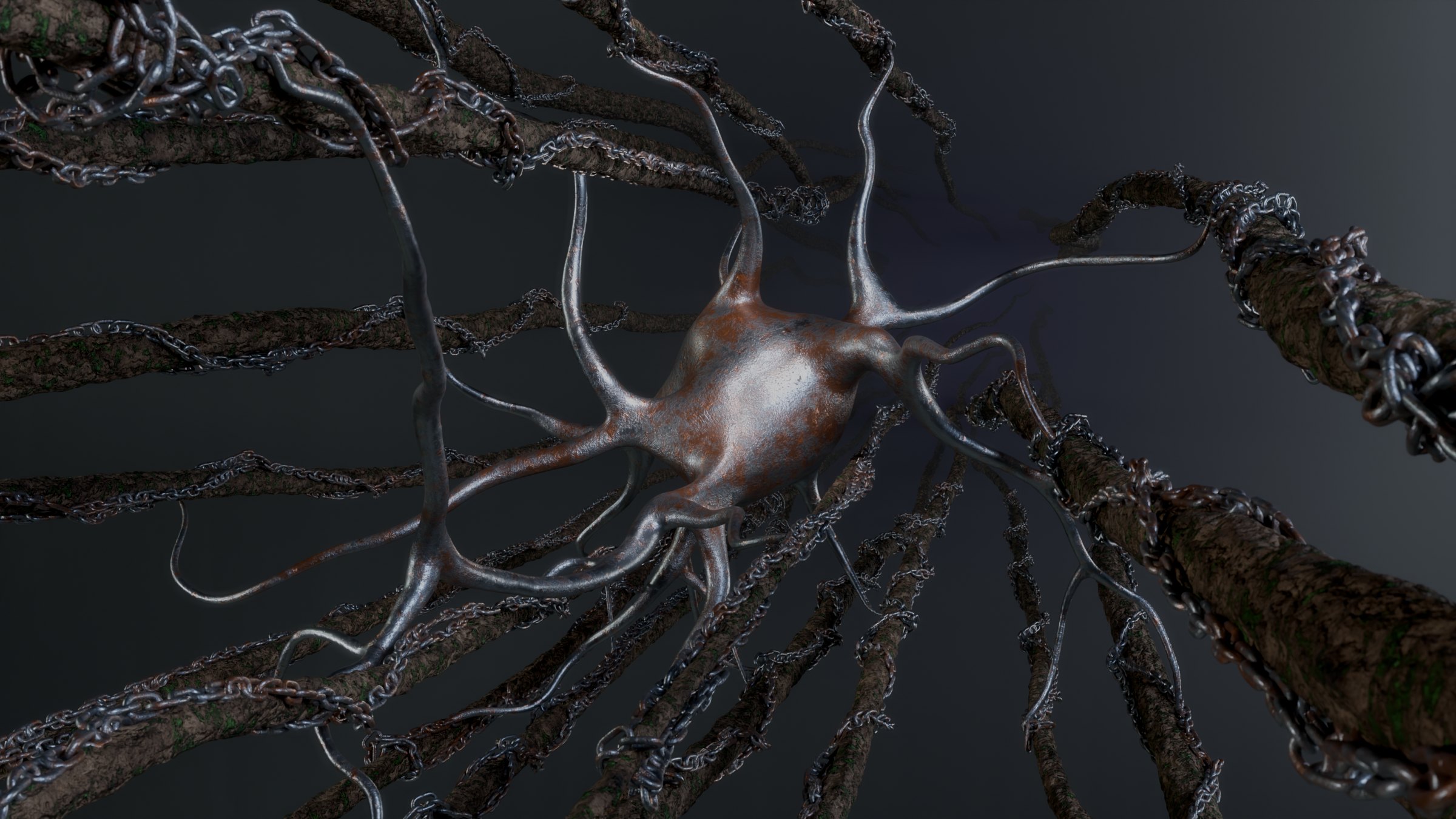
Learning to juggle or to speak a new language physically reshapes the brain's white matter. Myelin isn't as static as people once thought — sheaths lengthen and shorten across life, and when neighbors are lost to injury or age, surviving sheaths can stretch to fill the gaps. But how a cell decides how much myelin to make, and where to stop, remains poorly understood.
We're dissecting the cytoskeletal machinery that sets sheath length, using live imaging in zebrafish alongside genetic screens. We then ask whether the same machinery is redeployed in the adult mouse cortex during learning and in the compensatory growth that follows sheath loss. Beyond mechanism, these tools let us tune sheath dimensions deliberately, allowing us to perform the causal experiments in Direction 3.
The brain doesn't myelinate every axon uniformly. Instead, myelin is distributed in selective, patterned ways, especially along the fast-spiking interneurons that keep cortical rhythms in sync. Why myelin is arranged this way, and what happens to a circuit when that arrangement is disrupted, is among the biggest open questions in the field. We're especially interested in the branch points where axons split and signals can fail and whether the precise placement of myelin there helps neurons fire in time with one another.
Progress here has been limited by a hard problem: it's difficult to alter myelination patterns without first changing neuronal activity. We've developed ways to manipulate specific features of myelin placement independently of activity, letting us ask directly how those patterns support the fidelity of signal propagation across branch points. Paired with calcium or voltage imaging in neurons, this lets us link myelin geometry causally to neural activity, and to understand how its breakdown may contribute to disease and aging.
Together, these directions build toward a single goal: a mechanistic account of myelination that runs from the formation of single oligodendrocytes to the synchrony of a cortical circuit.
Interested in joining? →